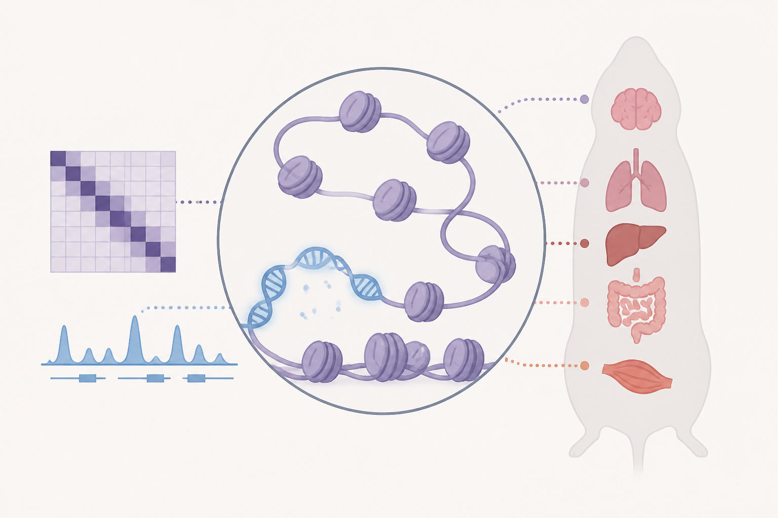
AI Generated Image
Spatial chromatin architecture and accessibility co-profiling of mammalian tissues
Nature • • health
Key Points:
- Spatial-ATAC-Hi-C is a novel technology that spatially profiles 3D genome architecture and chromatin accessibility simultaneously in tissue sections, using barcoded oligonucleotides and microfluidic indexing to preserve spatial information at a pixel resolution of 2,500 per tissue slide.
- Benchmarking experiments demonstrated that Spatial-ATAC-Hi-C accurately captures chromatin interactions and accessibility comparable to bulk in situ Hi-C and ATAC-seq, with slight enrichment for open chromatin regions due to in situ Tn5 tagmentation.
- Application to adult mouse brain sections revealed spatially resolved, cell-type-specific chromatin compartments, loops, and accessibility patterns that correspond well with known anatomical regions and single-cell ATAC-seq data, identifying distinct excitatory, inhibitory, and non-neuronal cell populations.
- Spatial-ATAC-Hi-C was successfully applied to human brain cerebellum and glioblastoma tumor samples, uncovering spatial heterogeneity in chromatin architecture and copy number variations (CNVs), including known oncogenic amplifications and structural variants validated by whole-genome sequencing.
- The technology shows promise for clinical applications by enabling spatially resolved epigenomic characterization of complex tissues and tumors, providing insights into tumor heterogeneity, 3D genome organization, and regulatory mechanisms in health and disease.


